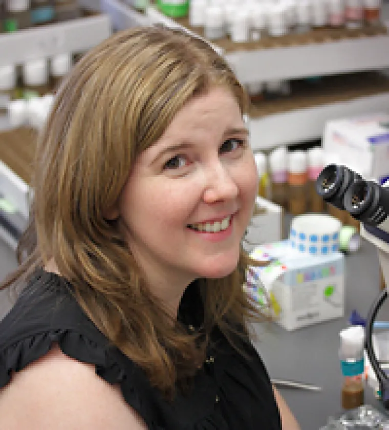
Megan Corty, Ph.D.

Documents
The Corty lab studies HOW and WHY glia wrap axons using Drosophila melanogaster (fruit flies) as our model organism.
Many axons in our brains and ALL of the axons in our peripheral nerves are ensheathed by specialized glia cells. We know these glia cells shape and support the physiology, structure, function, and health of the axons they wrap, but we don’t understand precisely how they do this. Knowledge of how this close relationship between glia and their axons develops, and precisely how they provide their supportive functions will hopefully allow us to do things like replace or substitute for missing or damaged glia and increase their supportive functions to help sick or injured neurons recover.
We study the wrapping glia in Drosophila peripheral nerves as a model of non-myelinating ensheathment because they are genetically accessible and allow us to probe neuron-glia interactions in vivo (i.e. in a live, functioning animal as opposed to a culture system). We use genetic techniques and imaging to conduct our research.
Biography: Dr. Corty received her B.A. in Human Biology from Stanford University before heading east to earn her Ph.D. in Neurobiology from Columbia University, where she studied dendrite morphogenesis using Drosophila as a model system in the lab of Wesley Grueber. Her love of glia began in her undergraduate years at Stanford where Ben Barres was pioneering and championing the study of these oft-overlooked brain cells. Though she focused on neurons for her graduate work, she was determined to study glia one day. She also fell in love with Drosophila as a model system because of how easily you could modify gene expression and visualize cells in vivo. The combination of glia and Drosophila led her to the lab of Marc Freeman for her postdoctoral work, where she began studying axon-glia interactions in the fly.

